Ophthalmology · Adelaide & Victor Harbor
Specialist EyeOPHTHALMOLOGY
MACULA & MEDICAL RETINA

Macular &
medical retinal care.

Care for disease of the macula (the central retina used for detailed vision) and the wider retina (the light-sensitive lining at the back of the eye). The choice is guided by the condition, retinal imaging, response and your wider health.

Wet age-related macular degeneration (AMD)+

Abnormal vessels beneath the retina can leak fluid or bleed, affecting central vision. Treatment aims to control activity and preserve sight. VEGF is a protein that can drive abnormal vessel growth and leakage. Treatment may block VEGF alone or both VEGF and angiopoietin-2, another protein involved in unstable blood vessels.

Imaging and response guide the choice and timing. Treatment is usually ongoing, and a longer interval is appropriate only when the eye remains controlled.

Dry AMD & geographic atrophy+

Age-related macular degeneration (AMD) can cause dry changes, ranging from early signs to geographic atrophy: areas where cells of the central retina and its supporting layer are lost. Assessment includes the location and growth of atrophy and whether wet AMD is also present.

For selected geographic atrophy, treatment targeting complement, part of the immune system, may be considered to slow growth of the atrophic area. It does not restore lost sight, is not suitable for all dry AMD and requires a careful discussion of treatment burden and risks, including inflammation and new wet AMD. Suitability, access and cost need individual discussion.

Smoking cessation, appropriate nutrition, selected use of AREDS2 vitamin and mineral supplements and support for low vision also matter. Supplements are not indicated for everyone; ask whether they suit your stage of AMD.

Diabetic eye disease+

Care can include observation, intravitreal injections, steroid implants, laser or surgery. Macular swelling and abnormal new vessels need different treatment plans. Blood glucose, blood pressure, cholesterol and general medical care remain important alongside eye treatment.

Retinal vein occlusion+

A blocked retinal vein can lead to macular swelling, haemorrhage (bleeding) and retinal ischaemia (reduced blood supply). Treatment may include targeted injections or a steroid implant for selected eyes. Laser may be needed for abnormal new vessels. Monitoring remains important even when swelling improves.

Central serous chorioretinopathy (fluid beneath the macula)+

Fluid beneath the macula has several possible causes. Some central serous cases can be observed; persistent disease may require treatment such as photodynamic therapy (PDT), which combines a light-sensitive medicine with a specific laser, after specialist assessment. Steroid exposure is reviewed with the prescribing clinician rather than stopped without advice.

Diagnosis matters: not every retinal fluid collection needs the same injection. Treatment or onward referral is arranged according to the findings and available services.

More than one
treatment pathway.

Treatment is matched to the diagnosis. A change in approach can be appropriate if the response, risks or burden change.

01

Controlling abnormal leakage

Some treatments target VEGF, a signal involved in abnormal blood vessel growth and leakage. Dual-pathway treatment also targets angiopoietin-2 (Ang-2), a protein involved in vessel stability. Selection depends on the diagnosis and response.

02

Inflammation

Steroid injections or implants that release medicine gradually can treat selected macular oedema (swelling of the central retina) and inflammatory conditions. Eye pressure and cataract risk require monitoring.

03

Geographic atrophy

Selected eyes may be assessed for treatment targeting complement, part of the immune system. The aim is to slow the enlargement of damaged areas, rather than recover vision already lost.

04

Laser & surgery

Retinal laser, light-activated treatment called photodynamic therapy (PDT), or surgery involving the vitreous and retina may be appropriate for specific diagnoses. Observation is also an active management choice.

This is an overview of treatment categories, not a recommendation for a particular medicine. Your consultation establishes suitability and whether treatment or referral is appropriate.

Imaging that guides decisions

Optical coherence tomography (OCT) is a light-based scan that shows the layers of the retina and helps track swelling and other changes. OCT angiography (OCTA) uses the scan to map blood flow without injected dye. Dye tests called fluorescein angiography and indocyanine green angiography can show leakage and circulation when needed. Fundus autofluorescence (FAF) is imaging of natural signals from the back of the eye that can help assess the retinal supporting layer and areas of cell loss.

Tests are selected for the question being answered. A scan is interpreted alongside your examination and symptoms.

READING AN OCT SCAN

A cross-section of the macula.

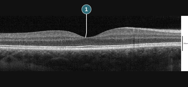
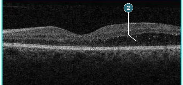
OCT uses light to show the retina in layers. These two examples help explain what your ophthalmologist looks for.

Normal macular example

Normal macular OCT, with number 1 marking the central foveal dip.Enlarge scan ↗
1 The normal central dip

The fovea is the small depression at the centre of the macula. Layers of retinal tissue are visible across the scan.

Macular oedema example

OCT showing diabetic macular oedema, with number 2 marking a dark fluid space within the retina.Enlarge scan ↗
2 Fluid within the retina

Dark spaces in this scan represent fluid within an area of swollen retinal tissue. This example is associated with diabetes.

Different eyes and scan settings; not a before-and-after comparison and not shown at a matched measurement scale. A scan alone does not establish the diagnosis or determine treatment. Your examination, symptoms and previous scans also matter.

Image sources & licences +

Normal scan: Wies6014, SD-OCT Macula Cross-Section, CC BY-SA 4.0. Number and leader line added; framed for display. Annotated adaptation shared under CC BY-SA 4.0.

Oedema scan: Jmarchn, Macular edema LE Man 61years Diabetic, CC BY-SA 3.0. One B-scan framed from the original image; number and leader line added. Annotated adaptation shared under CC BY-SA 3.0. These are external educational examples, not practice patient results.

Monitoring & treatment intervals

Some conditions need observation; others need repeated treatment even when vision seems stable. With a treat-and-extend plan, treatment is given and the gap to the next visit is adjusted according to disease activity, where suitable. Longer intervals are not guaranteed and are not appropriate for every condition.

Bring questions about discomfort, cost, transport or the visit schedule to your review. Do not stop or delay planned treatment without discussing the plan with your treating team.

Low-vision services, magnification, better lighting and practical support can help when sight remains affected despite treatment.

What to expect after an injection →
Intravitreal injection appointments

Read what happens at an injection visit, how follow-up is planned and what to ask before treatment.

Intravitreal injection information ↗
CONTACT THE PRACTICE

Contact the practice.

For appointments, referrals or a question about your care, speak with our practice team.

08 7070 2502 ↗Find your clinic →